For our international customers, please be advised that orders cannot be placed through our website by customers in countries with International Distributor representation.
Hexokinase - Manual
Source:
Yeast
CAS:
9001-51-8
EC:
2.7.1.1
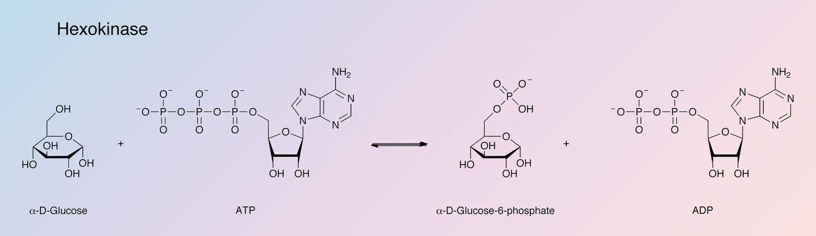
Hexokinase catalyzes the reaction:
Hexokinases have been isolated from the yeast cell in two distinct forms, designated P-I and P-II (Schulze et al. 1969). These are separate, noninterconvertible isozymes (Womack et al. 1973).
Hexokinase is used to determine glucose, fructose, mannose and ATP.
Characteristics of Hexokinase from Yeast:
Specificity
The enzyme phosphorylates D-fructose, 5-keto-D-fructose (Avigrad et al. 1968), D-glucose, 2-deoxy-D-glucose, D-mannose and D-glucosamine. ATP and ITP have been demonstrated to transphosphorylate in the yeast hexokinase reaction (Martinez 1961). The activity of P-I with fructose is 2.6 times that with glucose, whereas with P-II, a fructose:glucose ratio of 1:3 is obtained (Lazarus et al. 1966). The substrate specificities of yeast hexokinase have been extensively studied by Bessell et al. (1972).
Composition
Both P-I and P-II contain the same amino terminus, valine, and the same carboxy terminus, alanine. Amino acid composition has been reported by Schmidt et al. (1973b).
Characteristics of Hexokinase
Molecular Weight
The native forms have molecular weights of about 100,000 (Schulze et al. 1969) and consist of polypeptide chains of molecular weights slightly higher than 50,000 (Schmidt et al. 1973).
Optimal pH
7.5 - 9.0 (Sols et al. 1958).
Isoelectric point
P-I, 5.25 and P-II, 4.93 (Schmidt et al. 1973).
Extinction Coefficient
![]() = 8.85 for P-I and 9.47 for P-II (Schmidt et al. 1973).
= 8.85 for P-I and 9.47 for P-II (Schmidt et al. 1973).
Activators
Hexokinase requires magnesium ions for its catalytic activity. It is activated by catecholamines and related compounds (Harrison et al. 1972). Calcium ions do not affect the enzymatic activity.
Inhibitors
The enzyme is inhibited by compounds which react with SH groups. It is also inhibited by sorbose-1-phosphate, polyphosphates, 6-deoxy-6-fluoroglucose, 2-C-hydroxy-methylglucose, xylose and lyxose (Sols et al. 1958 and McDonald 1955).
Stability
Both the lyophilized preparation and the crystalline suspension are stable for 6-12 months at 2-8°C.
Hexokinase Products
Description
Activity
Code
Cat. #
Size
Price
Description
Hexokinase, Lyophilized, Recomb
Source:
Recombinant Hexokinase from Yeast
A lyophilized powder. Phosphohexose isomerase, 6-phosphogluconate dehydrogenase, adenylate kinase and creatine phosphokinase contaminant activities are 0.10%, 0.005%, 0.005% and 0.005%, respectively.
Store at -20°C.
Code
HKQLR
Product details
LS002515
2.5 ku
$69.00
LS002516
10 ku
$230.00
LS002518
Bulk
---
Description
Hexokinase, Lyophilized
Source:
Yeast
Chromatographically purified. A lyophilized powder containing ~30% buffer/stabilizer. Phosphohexose isomerase, 6-phosphogluconate dehydrogenase, adenylate kinase and creatine phosphokinase contaminant activities are 0.10%, 0.005%, 0.005% and 0.005%, respectively.
Store at -20°C.
Code
HKQL
Description
Hexokinase, Suspension
Source:
Yeast
Chromatographically purified. A suspension in 2.8M ammonium sulfate. Phosphohexose isomerase, 6-phosphogluconate dehydrogenase, adenylate kinase and creatine phosphokinase contaminant activities are 0.05%, 0.003%, 0.005% and 0.005%, respectively.
Store at 2-8°C.
Code
HKQS